research

Measuring organ function with CT

I develop quantitative CT imaging methods that combine tracer-kinetic modelling with physics-informed deep learning to measure lung and heart function for safer radiotherapy.

Research interests

Physics-informed AI

Physics- and physiology-informed AI to improve pharmacokinetic modelling.

Cardiopulmonary imaging

Functional imaging of cardiopulmonary physiology.

Personalized cancer treatment

Using functional imaging in personalized cancer treatment, including functional avoidance radiotherapy planning to spare healthy tissue and reduce toxicity.

01

Lung ventilation, lung perfusion and myocardial perfusion from a single dynamic contrast-enhanced CT scan PhD thesis
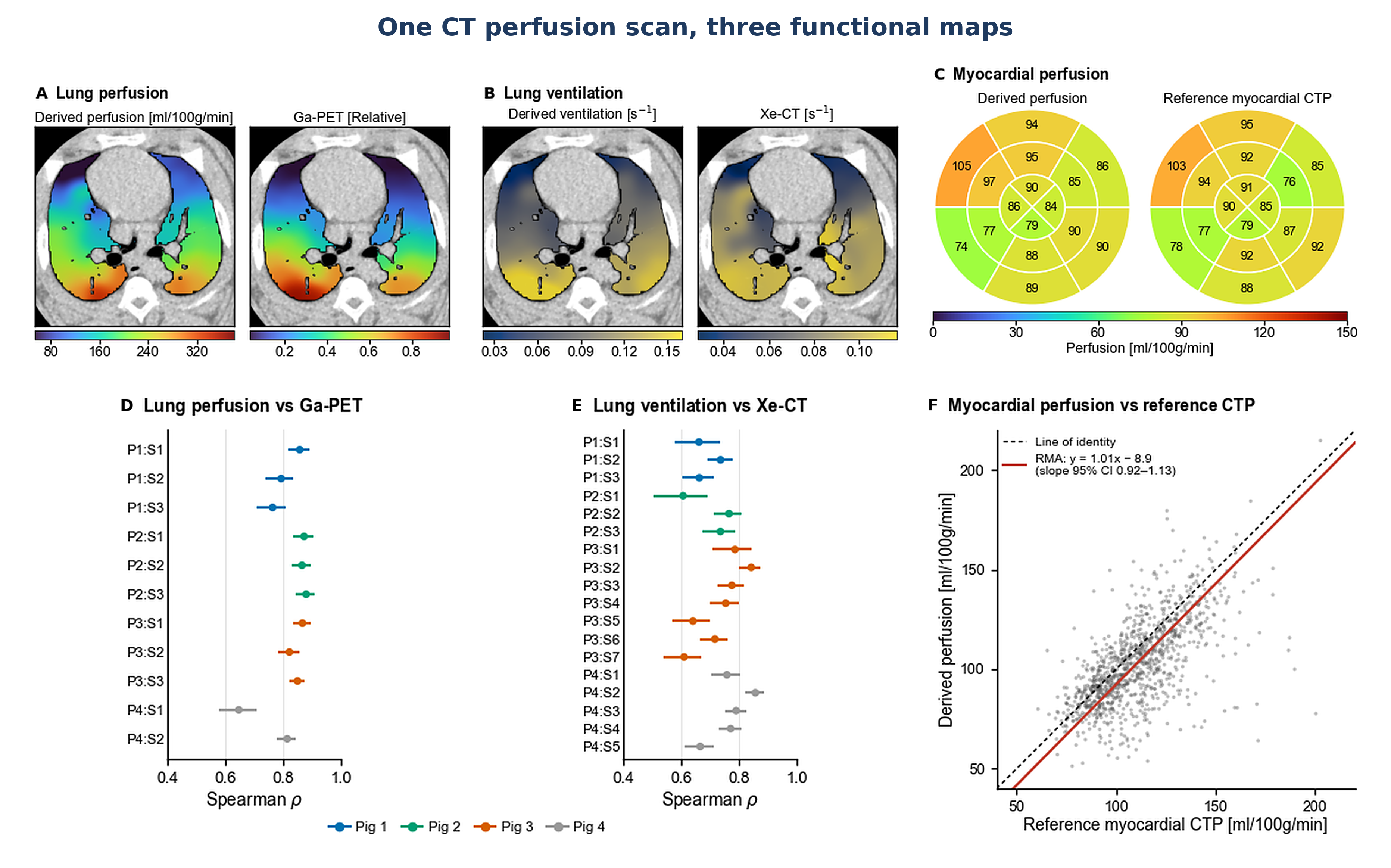
One CT perfusion scan, three functional maps: derived lung perfusion, lung ventilation and myocardial perfusion maps beside Ga-PET, Xe-CT and reference myocardial CT perfusion, with per-scan Spearman correlations and a regression of myocardial perfusion against reference CTP. Full size
(A–C) Derived lung perfusion, lung ventilation and myocardial perfusion vs. reference standards; (D–F) their agreement across scans and myocardial segments.

Functional lung avoidance radiotherapy aims to protect the best-working parts of the lung during lung cancer treatment, and the heart is an equally important organ at risk. Mapping lung ventilation, lung perfusion and heart muscle perfusion would normally take three separate nuclear medicine scans with different tracers.

This project derives all three from a single dynamic contrast-enhanced CT scan acquired in the same session as the radiotherapy planning CT. Deformable registration and tracer-kinetic modelling separate the breathing and blood-flow signals in the lung, and the heart is reconstructed at mid-diastole to measure myocardial perfusion.

Repeated at follow-up, the same scan could also help catch early lung and heart toxicity after treatment.

Deformable registration Tracer-kinetic modelling CT perfusion Functional lung avoidance RT Cardiac toxicity Pulmonary embolism
Key results In animal studies, all three maps agreed closely with established reference standards:
  • Lung ventilation vs. xenon-enhanced CT: pooled Spearman ρ = 0.73 (per-scan 0.61–0.86).
  • Lung perfusion vs. 68Ga-MAA PET: pooled ρ = 0.83 (per-scan 0.65–0.88).
  • Myocardial perfusion vs. conventional cardiac CT perfusion across the 16 AHA segments: regression slope 1.01 (95% CI 0.92–1.13).
Related
02

Myocardial perfusion from multiphase coronary CTA

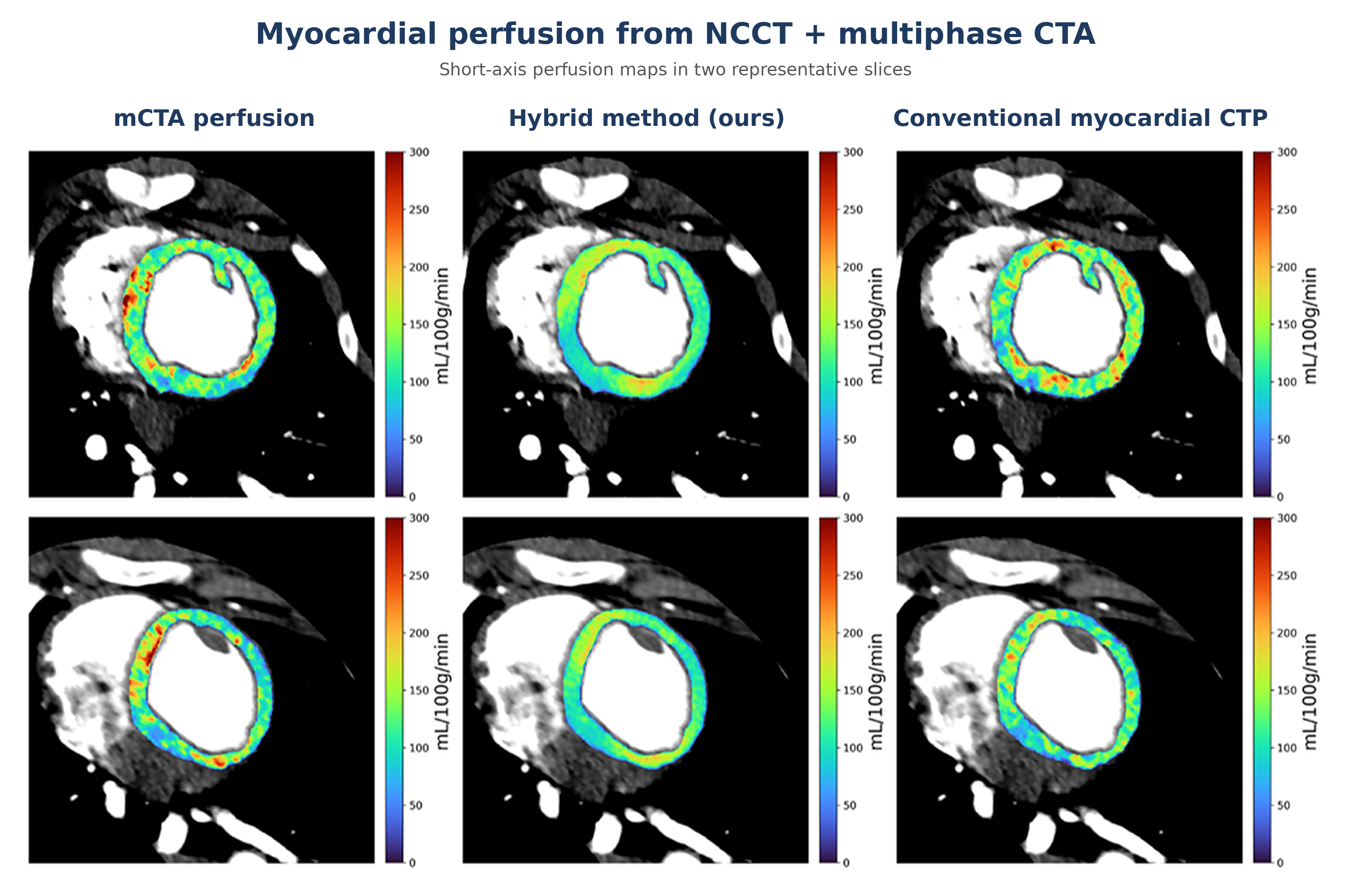
Short-axis myocardial perfusion maps in two representative slices, comparing mCTA perfusion, the hybrid method, and conventional myocardial CT perfusion. Full size
Short-axis myocardial perfusion maps in two representative slices: mCTA perfusion, the hybrid method, and conventional myocardial CTP.

Coronary CT angiography (CTA) shows where the coronary arteries are narrowed, but not whether the heart muscle is getting enough blood. That usually requires a separate CT perfusion scan, which adds time and radiation dose.

This project turns a standard coronary CTA into a combined anatomical and functional exam. Two short delayed phases are added to the routine CTA, and together with a non-contrast CT they let a tracer-kinetic model estimate myocardial blood flow. Because so few time points make these estimates noisy, a residual U-Net then refines the perfusion maps.

The aim is wider access to stenosis and ischemia assessment from a single, lower-dose study.

Tracer-kinetic modelling Residual U-Net Coronary CTA Radiation cardiotoxicity Myocardial ischemia
Key results In 12 animals imaged weekly (77 exams, leave-one-animal-out cross-validation), refinement cut the segment-level error against reference CT perfusion from 18.86 to 13.58 mL/100 g/min, a 28% reduction (p < 0.001).
  • Limits of agreement narrowed by 31%.
  • Bias fell from +6.93 to −1.09 mL/100 g/min.
  • Structural similarity rose from 0.761 to 0.795.
  • The reference scan itself carries up to about 7.8 mL/100 g/min of measurement error, so the refined maps are approaching the best agreement possible.
Related
03

Stroke: brain perfusion and thrombectomy eligibility from multiphase CTA In progress

This project generates brain perfusion maps from multiphase CT angiography (mCTA) and uses AI to segment the ischemic core and penumbra. The segmented volumes are then used to apply the DEFUSE 3 criteria and assess whether a stroke patient is eligible for endovascular thrombectomy (EVT).

Multiphase CTA Perfusion mapping AI segmentation DEFUSE 3 Endovascular thrombectomy

No publications yet.

Full publication list on the publications page and Google Scholar; conference presentations on the talks page.